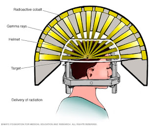
The word knife is a misnomer, this is basically a non-invasive (non-surgical) technique which uses intersecting Gamma rays emitted from a radiological material usually Cobalt 60, the technique is also called stereotactic radiosurgery.
The knife has an associates specialized software program which helps the doctor to locate and irradiate small targets in the head (brain), blood vessels and other tissue with extreme precision. An intense dose of the radiation can be given on the target area leaving the normal surrounding tissue unharmed.
You might confuse Gamma Knife with CyberKnife, but each one has different approaches, mainly a Gamma Knife targets the brain and cervical spine with mainly one treatment session while a CyberKnife can treat brain cancer in almost all body parts in about 5 treatment sessions.
Gamma Knife Working Mechanism
 |
| Gamma Knife Banned |
Gamma knife utilized multiple beams which are converged in a three-dimensional plane to focus on the tissue to be eradicated, Gamma Knife has 2 modules:
A) Perfexion Model
B) Icon model
Perfexion model
It consists of 192 sources of cobalt 60 attached to the source core each of which produces a fine beam of Gamma rays. The sources are evenly distributed over the source core so that the beam is directed at a common center. The resultant intensity at the center of the focus , where the tumor will be most likely a brain tumor, is very high but the intensity a little away from the center is very low due to which the nearby sensitive and healthy tissues are spared.
The rays produced are ellipsoid in shape and by overlapping multiple foci of radiation, the shape for the type of abnormality can be exacted and brain tumor removal surgery can be planned accordingly by several doctors.
Sensitive structure of the brain such as spinal cord,globus pallidus, hypothalamus, lenticular nucleus, putamen, optic chiasma, etc.are avoided and their safety is ensured.
Procedure for Perfexion model
The procedure is done in a single session, under local anesthesia contrary to the general anesthesia used in many procedures. A rigid head frame incorporating three dimensional systems is attached to the patients’ skull with four screws. The radio-imaging reports using Magnetic Resonance Imaging (MRI), computerized tomography (CT), or angiography are sent to the knife’s planning computer system and various doctors program the procedure keeping in mind the normal human anatomy, the abnormality and the variations of anatomy in different individuals.
The targeted brain tumor is then placed in the center approximately 200 precision-aimers, converging the beams of cobalt 60 generated Gamma rays. The time and dose intensity depends on the shape and size of the abnormal tumor tissue that need to be targeted.
Icon model
Multiple sessions are generally required in the Icon model and the external frame is not used contrary to Perfexionmodel which consists of a single session and an external frame on the patients' skull.
 |
| External Frame |
Diseases which can be treated using gamma knife include:
- Malignant tumors of both primary and secondary origin in the brain.
- Benign tumors of the brain such as meningioma pituitary adenoma etc.
- Blood vessel defects such as AV malformations AVM.
- Trigeminal neuralgia.
Precautions
Since the procedure requires a great precision so these things are kept in mind before and during the surgery:
● Rigid attachment of head frame.
● The accuracy of imaging studies.
● Shaping the tissue to be treated.
● The precision of attachment of Gamma Knife to head unit.

No comments:
Post a Comment
We are happy that you want to comment, please note that your comment will be reviewed first before it is published.
If you like the article! You can share it with your friends and colleagues by pressing at social media buttons provided to the left of the page.
NO word verification or sign up is required!