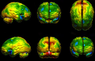
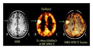
SPECT or Single Photon Emission Computerized Tomography also called SPET, in short, is an imaging technique using gamma rays and provides a three-dimensional information about the flow the blood to different organs of the body. It is usually presented as cross-sectional slices through the patient but can be freely reformatted or manipulated as required.
How a SPECT Machine Works
The patient is injected with radioactive isotopes like iodine-123, technetium-99m, xenon-133, thallium-201, and fluorine-18 these materials stay in the bloodstream and produce gamma rays which are detected by the scanner.
The type of tracer used depends on what your doctor wants to measure. For example, if the doctor is looking at a tumor, he might use radioactive glucose called Fluorodeoxyglucose (FDG) and watch how it is metabolized by the tumor. The test differs from pet scan as pet scan uses a material which is absorbed by the tissue and the working of tissue can be seen but here tracer is such a material which is not absorbed but rather retained inside the bloodstream so that the amount of perfusion can be seen. The material used here have comparatively longer half-life for example, technetium-99m (t½ = 6 hours), indium-111 (t½ = 2.8 days), iodine-123 (t½ = 13.22 hours) and thallium-201 (t½ = 73 hours).
 |
| NueroSPECT |
Scan Procedure of a SPECT
The patient is injected in the medial cubital vein usually with the tracer element and is asked to rest for 10 to 20 minutes until the tracer element has reached the part of the body to be scanned usually brain.
The patient is then scanned and the images produce help in the diagnosis and plan the future treatment of the patient. For example Pictures from a scan may have colors that highlight to the doctor which areas of the body absorbed more radioactive tracer and which ones absorbed less, making the total scan time be around 15 to 20 minutes.
The patient is then scanned and the images produce help in the diagnosis and plan the future treatment of the patient. For example Pictures from a scan may have colors that highlight to the doctor which areas of the body absorbed more radioactive tracer and which ones absorbed less, making the total scan time be around 15 to 20 minutes.
After the procedure, the doctor usually advises increasing the fluid intake to wash out the tracer element from the body easily.
Where is the Technology Used
Brain imaging -Currently, SPECT and SPECT-CT imaging studies of the brain play a primary role in patient diagnosis. The various diseases which can be evaluated are :
- Dementia
- Clogged blood vessels
- Seizures
- Epilepsy
- Head injuries
Myocardial Perfusion Scan - This technique evaluates heart conditions such as coronary artery disease (CAD) and the motion of heart chambers.Technetium-99m tetrofosmin is the isotope used. Following this, the heart rate is increased to induce myocardial stress, either by exercise on a treadmill or pharmacologically with drugs such as adenosine, dobutamine, or dipyridamole. This helps to the blood flow to the different areas of the heart. MPI has been demonstrated to have an overall accuracy of about 83% (sensitivity: 85%; specificity: 72%)
Bone - This is used to diagnose hidden bone fracture and bone healing
Some Risks of a SPECT Scan
It includes various risks like:
The patient receiving the tracer can be allergic to the tracer.
Patients with bleeding disorders may bleed a lot when injected with the tracer.
The tracer can act cross placenta and act as a teratogen in pregnant females.
The radiations can cause mutations in few individuals.


No comments:
Post a Comment
We are happy that you want to comment, please note that your comment will be reviewed first before it is published.
If you like the article! You can share it with your friends and colleagues by pressing at social media buttons provided to the left of the page.
NO word verification or sign up is required!