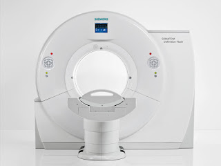
What Is a CT Scanner?
Computerized tomography (CT or commonly called CAT) is a technique which uses x rays to take images in multiple planes and these images are then assembled in a computer to form a three-dimensional picture of the organ to be viewed. This type of special X-ray, in a sense, takes "pictures" of slices of the body so doctors can look right at the area of interest.
Computerized tomography (CT) provides good details for soft organs and blood vessels compared to normal x rays. Using this technology the radiologist can interpret the cancers, trauma, vascular injuries, musculoskeletal deformities, etc in an easy and precise way.
 |
| CT Scanner of Donut Like Shape |
Working Principle of the CT Scanner
CT is based on the fundamental principle that the density of the tissue passed by the X-ray beam can be measured from the calculation of the attenuation coefficient. Using this principle, CT allows the reconstruction of the density of the body, by two-dimensional section perpendicular to the axis of the acquisition system.
The CT X-ray tube emits N photons (monochromatic) per unit of time. The emitted X-rays from a beam which passes through the biological later of thickness delta x.A detector placed at the exit of the sample, measures N + delta N photons, delta N smaller than 0.
There are two processes of the absorption:
- Photoelectric effect
- Compton effect
The phenomena are represented by the coefficient mu.
Unlike x-ray which uses a fixed x-ray tube CT scan uses a revolving X-ray tube that rotates around the circular opening of a donut-shaped structure called a gantry. During the CT scan patient, lies on a bed that slowly moves inside the gantry while the mobile X-ray tube rotates around the patient shooting x-rays at the patient's body and contrary to X-ray which uses a plate CT uses X-ray detectors which detect the x-rays coming from the body part under examination.
Inside the CT Scanner
How a CT Image is Created?
Each time the tube completes around the computer forms a two-dimensional image of the body part just like a slice of the part has been cut off and presented. Multiple slices of the part under examination are prepared.
The thickness of each slice varies but is usually between 1-10mm in thickness and the number of slices varies on the pathology to be detected. Once all the slices are taken the computer arrange them together using an algorithm and forms a three-dimensional image of the organ.
The three-dimensional image formed can be rotated and different slices can be viewed individually and the pathology can be identified.
 |
| Effective Radiation Doses For Adults |
Uses of CT Scanner
1)A CT Scanner is the fastest and most accurate tools for examining the chest, abdomen, and pelvis.
2) CT Scanner is used to diagnose various tumors including:
- Breast cancers
- Abdominal tumors
- Lymphoma
- Lung cancer
- Renal cancer
- Hepatic cell carcinoma
- Osteosarcoma
3) CT Scanner is used to examine patients with injuries and trauma
4) CT Scanner is used to diagnose radiopaque masses in various organs which include:
- Renal stone
- Ureteric stone
- Gallstone containing calcium phosphate and are radiopaque
5) CT Scanner can also help in the diagnosis of vascular diseases including:
- Stroke
- Thromboembolism
- Deep venous thrombosis
- Transient ischemic attack

No comments:
Post a Comment
We are happy that you want to comment, please note that your comment will be reviewed first before it is published.
If you like the article! You can share it with your friends and colleagues by pressing at social media buttons provided to the left of the page.
NO word verification or sign up is required!