INTRODUCTION
Lasers have revolutionized every specialty of medicine since its invention almost half a century back. In 1961, the ruby laser was the first one to have found clinical application in ophthalmology. Presently, lasers have become an indispensable tool in diagnostics and therapeutics for a wide spectrum of diseases, involving both anterior and posterior segments of the eye like glaucoma, cataract, diabetic retinopathy, etc.
CLINICALLY AVAILABLE LASERS IN OPHTHALMOLOGY
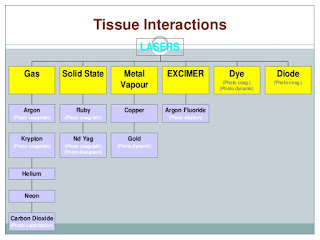
The results of the attempting to instrumentalize sunlight in ophthalmic surgery by the famous German ophthalmologist, Dr. Gerhard Meyer Schwickerath changed medical history and laid the foundation for modern laser surgery as we know it today. Today, a large variety of different lasers are used for surgery and therapy in ophthalmology. Some of the commonly available lasers and their clinical applications are listed below:
 |
| Role of Lasers in Ophthalmology |
Excimer Laser: It is a Photoablative laser with 193nm wavelength and is used in epithelial and anterior stromal keratopathies, PRK and LASIK. It falls under the Ultra-violet spectrum.
The excimer laser is mainly used in vision correction of myopia, astigmatism or hyperopia. There are certain risks related to doing such eye surgery, you can see an article published by FDA to reduce such risks.
The excimer laser is mainly used in vision correction of myopia, astigmatism or hyperopia. There are certain risks related to doing such eye surgery, you can see an article published by FDA to reduce such risks.
Argon laser: It is a blue-green or green laser with working wavelength of 488-514nm. It acts by photocoagulation and is used in iridotomy, trabeculoplasty, sclerostom, etc
Krypton Diode: This is also a photocoagulation laser with 647nm wavelength. Used in Retinal photocoagulation.
Nd: YAG - Continuous wave: It is an infrared laser used in capsulotomy, cyclo-photocoagulation and cataract surgery.
Nd: YAG - Q-switched or mode-locked: Acts on the principle of photo-disruption, finds application in Retinal photocoagulation, iridotomy, trabeculoplasty, sclerostomy, hyaloidotomy, etc
Ho: YAG: Infrared laser, working on photoablation principle, used for thermo-keratoplasty, sclerostomy, etc.
Er: YAG: Also an infrared laser with 2940nm wavelength. It causes photoablation and is used for skin resurfacing, cataract surgery, sclerostomy etc.
CO2: It has a wavelength of 10,600nm and is used in blepharoplasty, skin resurfacing, conjunctival carcinoma in situ, etc.
DIAGNOSTIC APPLICATIONS OF LASERS
In the field of diagnostics, lasers find applications primarily due to the development of confocal scanning laser ophthalmoscope (cSLO) and optical coherence tomography (OCT).
The cSLO generated high contrast retinal images by means of laser light of different wavelength without dilation of the pupil. The cross-sectional view of ocular structures, especially the retina, with high depth resolution using low-coherence light sources can be obtained by OCT.
Imaging of anterior segment
OCT can be used to view cross-section of the cornea and complex anatomy as it is a dehydrated tissue and transparent. The cellular characteristics of corneal structures can be visualized with cSLO.
 |
| Retinal OCT Image |
The Figure shows multispectral cSLO plus OCT allows for multimodal evaluation of retinal abnormalities. Different wavelengths of multispectral cSLO reveal different features of complex lesions, such as choroidal neovascular membranes. The OCT device becomes a more effective directed study when guided by cSLO.
In glaucoma diagnosis
Glaucomas are the leading cause of blindness in the world1. OCT and Auto-fluorescence can help in early detection and diagnosis, thus helping in preventing blindness at a later stage.
Optical coherence tomography
Helps by obtaining accurate and reliable measurements of retinal nerve fiber layer thickness and early glaucoma diagnosis.
Autofluorescence
Lipofuscin in the retinal pigment epithelium derives mainly from incomplete degradation of outer photoreceptor segments. Histologic and spectroscopic results revealed an accumulation of lipofuscin as the dominant fundus fluorophore localized in the lysosomes of the retinal pigment epithelium in the parapapillary atrophic zone.
Scanning laser ophthalmoscopy enables the detection of fundus autofluorescence level and distribution in vivo with high resolution and reproducibility.
Increased autofluorescence properties have been reported in the parapapillary region in patients with ocular hypertension, primary and secondary open angle glaucoma. An example for this application is the Heidelberg Retina Angiograph HRA (Heidelberg Engineering), which uses an Argon- Blue-Laser (488nm) for excitation and a barrier filter for detection of emitted light above 500 nm.
 |
| Fundus Autofluorescence |
THERAPEUTIC APPLICATIONS OF LASERS
Oculoplastics
The CO2 laser is used for blepharoplasty, ablation of eyelid lesions, such as seborrheic keratosis, chalazia, adnexal tumors, and lipid plaques, ablation of conjunctival papillomas or melanosis, and skin resurfacing. The CO2 laser’s 0.1 mm depth of penetration allows precise removal of eyelid lesions involving the medial canthus and puncta. Its use in blepharoplasty involves skin and conjunctival incisions and excision or vaporization of fat.
Use of the CO2 laser results in significantly less bleeding and postoperative swelling and ecchymosis; however, healing takes longer because of sealing of blood vessels and heat necrosis.
Use of the CO2 laser results in significantly less bleeding and postoperative swelling and ecchymosis; however, healing takes longer because of sealing of blood vessels and heat necrosis.
Both the CO2 laser and the erbium: yttrium aluminum garnet (Er: YAG) laser are used for periocular resurfacing of wrinkles and blemishes. Laser resurfacing is believed to have less risk of scarring and dys-pigmentation because of the greater control of wound depth compared with dermabrasion and chemical peel.
Because significant thermal diffusion (and therefore thermal damage) will not occur if the pulse duration is less than the time it takes the tissue to cool, the super- pulsed and ultra-pulsed CO2 lasers were developed so that the pulses can correspond to the thermal relaxation time of the skin.
Because significant thermal diffusion (and therefore thermal damage) will not occur if the pulse duration is less than the time it takes the tissue to cool, the super- pulsed and ultra-pulsed CO2 lasers were developed so that the pulses can correspond to the thermal relaxation time of the skin.
Corneal surgery
The laser used commonly for corneal application is the excimer laser. Phototherapeutic keratectomy with the excimer laser has been used for corneal disorders involving the epithelium or anterior stroma along with refractive surgeries. The excimer laser is capable of reshaping the cornea by amounts of 0.5 lm with the collateral damage of less than 1 lm.
Laser in-situ keratomileusis (LASIK)
A microkeratome is used to create a corneal flap, hinged on one side, ablate the underlying stroma is ablated with an excimer laser and flap is replaced. LASIK has the benefit of reducing operative discomfort, corneal haze, and visual recovery time.
LASIK disadvantages are poor flap healing, epithelial growth under the flap and deeper stromal ablation. It can be used for hyperopia with large diameter corneal ablation.
LASIK disadvantages are poor flap healing, epithelial growth under the flap and deeper stromal ablation. It can be used for hyperopia with large diameter corneal ablation.
 |
| Flap Creation in LASIK Surgery |
The Figure Shows a schematic diagram of LASIK flap creation with raster pattern, while cornea is flattened by an applanating lens. (Right) Flap after lifting.
Other lasers that have been investigated for refractive surgery include the neodymium: Yttrium Lithium Fluoride (Nd: YLF), holmium: Yttrium-Aluminum-Garnet (Ho: YAG), Nd: YAG pumped optical parametric oscillator laser and a CW diode laser.
Glaucoma
Many lasers techniques have found application in the treatment of primary open angle glaucoma (POAG) and narrow-angle glaucomas.
Iridotomy and Iridoplasty
Laser iridotomy lasers are used in primary angle closure glaucoma, secondary pupillary block glaucoma, malignant glaucoma, plateau iris, etc. Different lasers can be used for this and each has its own advantage, eg: argon laser has better coagulative effects, Q-switched Nd: YAG laser has better long-term results.
Cataract Surgery
Nd: YAG and Er: YAG lasers are under clinical investigation for laser-assisted cataract surgery. The direct Nd: YAG system uses direct laser energy to fragment the lens while pulsed laser energy is used in indirect Nd: YAG system to strike a titanium target on the probe to generate optical breakdown and plasma formation. This creates shock waves which make contact with the lens material held in apposition to the probe tip by aspiration.
The Er: YAG system vaporizes the lens material by explosive vaporization forming a cavitation bubble and by micro-pulses traversing the cavitation bubble and generating energy.
Some advantages of laser-assisted cataract surgery are smaller incision size, minimal heat generated, reduced risk of damage to the posterior capsule and corneal endothelium, etc.
Other surgeries
Lasers have also found application in other treatments related to ophthalmology likeTrabeculoplasty, Trabecular ablation, Hyaloidotomy, Sclerostomy, Cyclophotocoagulation, Capsulotomy, Vitreoretinal disease, Diabetic retinopathy, Macular disease, Central and branch vein occlusion, Retinopathy of prematurity, Neoplasia, Retinal breaks and Nasolacrimal diseases, etc.
RECENT ADVANCES
Ultrafast Lasers in Ophthalmology
These lasers have become a promising tool for microsurgery in ophthalmology due to their precision. Their side effects are limited to the micrometer because of range low energy threshold.
IntraLase introduced its first commercial system in 2001 for replacing the mechanical microkeratome and since then these lasers have been used successfully in refractive surgery.
Schwind Eye Tech has the new TransPRK technique, where the laser automatically removes the epithelium layer and then applies refraction correction.
Femtosecond lasers in Ophthalmology
Though Femtosecond (FS) lasers are similar to Nd: YAG laser, they have ultra-short pulse duration, producing smaller shock waves and cavitation bubbles which affect a tissue volume about 103 times less. The first prototype was designed and constructed by Dr. Juhaszet al of in the early 1990s.
These lasers have found application predominantly in corneal flap creation in LASIK surgery under monikers as “IntraLASIK,” “all-laser LASIK,” and “bladeless LASIK.” Femtosecond Laser-Assisted Lamellar Keratoplasty, Femtosecond Laser–Assisted Penetrating Keratoplasty, Intracorneal Ring Segments and Femtosecond Laser–Assisted Astigmatic Keratotomy and Arcuate Wedge Resection are some of the other applications.
Two-photon technique
Multiphoton imaging combines diagnosis on a cellular level with the option of ablation in the micron range. In vivo multiphoton-autofluorescence, imaging and ablation by femtosecond laser pulses are possible from the outer face and may prove to be a clinically applicable novel option for minimal-invasive trabecular surgery.
Phacoemulsification of Opaque Lens Matter
Modern cataract surgery is centered around Phacoemulsification (ultrasound induced fragmentation) of opaque lens matter with preservation of the capsular bag. An alternative to the application of ultrasound pulses is the Dodick Laser Photolysis.
It applies a 1064 nm Nd: YAG-Laser pulse on a titanium target with the result of a shock wave at the tip, disrupting the lens nucleus without harming the thin capsular bag. A 1064 nm Q-switched Nd: YAG-laser with a pulse duration of 4 ns and an energy of 7 to 10 mJ is applied has also be used.
It applies a 1064 nm Nd: YAG-Laser pulse on a titanium target with the result of a shock wave at the tip, disrupting the lens nucleus without harming the thin capsular bag. A 1064 nm Q-switched Nd: YAG-laser with a pulse duration of 4 ns and an energy of 7 to 10 mJ is applied has also be used.
 |
| Phaco Cataract Removal |
CONCLUSION
Modern ophthalmic lasers have improved the diagnostic and therapeutic fronts in their evolution. Lasers have now become an indispensable tool in the armamentarium of ophthalmologists, but further research is needed for the development of cost-effective lasers and minimizing the adverse effects of the presently available ones.
Do you find this article informative? then please share it with your friends and colleagues. If you have any ideas related to this article you can post them in the comment section below. Did we miss any eye treatments using any type of laser?
Do you find this article informative? then please share it with your friends and colleagues. If you have any ideas related to this article you can post them in the comment section below. Did we miss any eye treatments using any type of laser?

No comments:
Post a Comment
We are happy that you want to comment, please note that your comment will be reviewed first before it is published.
If you like the article! You can share it with your friends and colleagues by pressing at social media buttons provided to the left of the page.
NO word verification or sign up is required!