Monday, March 26, 2018

TOP 7 Books In Biomedical Engineering


Here is a list of top 7 must-read books for anyone pursuing a course in Biomedical Engineering or having a keen interest in this field, or even working as a biomedical engineer in the medical domain and want to know more about various aspects of the biomedical field, you can make use of the Top 7 books in biomedical engineering field to advance and know more about various portions of the biomedical field. 


1.      3D Bioprinting and Nanotechnology in Tissue Engineering and Regenerative Medicine


3D Bioprinting and Nanotechnology in Tissue Engineering and Regenerative Medicine3D Bioprinting and Nanotechnology in Tissue Engineering is a comprehensive book highlighting the industrial applications of these technologies. It also covers other topics of current relevance such as nano-bio-materials and stem cells in tissue regeneration.
The book highlights the clinical applications, regulatory hurdles, and risk-benefit analysis of each technology and helps you in selecting the ideal materials and recognising the apt parameters for printing and incorporate biologically active agents and cells into a printed structure. The integration of 3D printing and nanotechnology is explained with an overview to improve the safety of application of these in aspects in biomedical applications.

The text also discusses legal and regulatory aspects and commercial overviews making it a perfect practical guide for anyone venturing in the field of biomedical technology by extrapolating theoretical aspects into an industrial or clinical setting. Application- wise sustainability and short-comings have been discussed for various technologies to make their commercial use feasible in meeting medical needs. The book brings a right blend of theoretical concepts and the latest principles and technologies.


Authors
Lijie Grace Zhang, John P Fisher, Kam Leong
Publisher
Academic Press, 2015
ISBN
0128006641, 9780128006641
Length
392 pages


Monday, February 12, 2018

Introducing Computerized Tomography

What Is a CT Scanner? 


Computerized tomography (CT or commonly called CAT) is a technique which uses x rays to take images in multiple planes and these images are then assembled in a computer to form a three-dimensional picture of the organ to be viewed. This type of special X-ray, in a sense, takes "pictures" of slices of the body so doctors can look right at the area of interest.

Computerized tomography (CT) provides good details for soft organs and blood vessels compared to normal x rays. Using this technology the radiologist can interpret the cancers, trauma, vascular injuries, musculoskeletal deformities, etc in an easy and precise way.

Saturday, February 10, 2018

Everything You Wanted to Know About Ophthalmology Laser Advances

INTRODUCTION


Lasers have revolutionized every specialty of medicine since its invention almost half a century back. In 1961, the ruby laser was the first one to have found clinical application in ophthalmology. Presently, lasers have become an indispensable tool in diagnostics and therapeutics for a wide spectrum of diseases, involving both anterior and posterior segments of the eye like glaucoma, cataract, diabetic retinopathy, etc.

CLINICALLY AVAILABLE LASERS IN OPHTHALMOLOGY


The results of the attempting to instrumentalize sunlight in ophthalmic surgery by the famous German ophthalmologist, Dr. Gerhard Meyer Schwickerath changed medical history and laid the foundation for modern laser surgery as we know it today. Today, a large variety of different lasers are used for surgery and therapy in ophthalmology. Some of the commonly available lasers and their clinical applications are listed below:

Role of Lasers in Ophthalmology


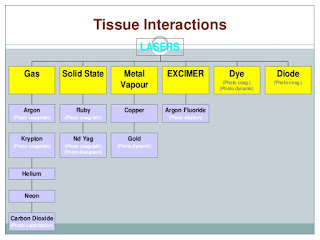
Excimer Laser: It is a Photoablative laser with 193nm wavelength and is used in epithelial and anterior stromal keratopathies, PRK and LASIK. It falls under the Ultra-violet spectrum. 

The excimer laser is mainly used in vision correction of myopia, astigmatism or hyperopia. There are certain risks related to doing such eye surgery, you can see an article published by FDA to reduce such risks

Wednesday, February 7, 2018

BILISPEC BILIRUBIN MEASUREMENT DEVICE

Jaundice is due to hyperbilirubinemia, which is particularly common in premature babies, who need an adequate liver capacity to discharge excess bilirubin. Affects approximately 60 percent of babies, can cause brain damage or even death if not treated.

In sub-Saharan Africa, where specialists often do not have access to expensive diagnostic equipment, babies are 100 times more likely to die from this cause than newborns in the United States.

Monday, February 5, 2018

All You Need To Know About Dermatology Laser Application Trends


INTRODUCTION


Over the past few decades, laser systems have been found extensive application in various clinical aspects of cosmetic surgery. From use in photocoagulation and vaporization of vascular malformations to removal of epithelial lesions, lasers have become an indispensable tool in surgery.

However, the thermal properties of laser treatment adversely affect the adjacent tissue, thus impacting wound healing and scar formation negatively. This problem has been addressed by the more selective and restrictive destruction of targeted tissue by the more recent laser systems like pulsed and Q-switched lasers matched to the target’s absorption characteristics.

These advances help in expanding the therapeutic spectrum of lasers in the field of cosmetic surgery and improvement in the quality of results achieved.

MODES OF OPERATION OF MEDICAL LASERS


Laser radiation used in dermatology work of the same basic principle but can be varied depending on the system used and the intended application keeping the three core variables in mind:


1. Wavelength


This can be tuned in dye lasers within a range by use of a single dye, the range can be broadened by exchanging the dye solution. Multiline regimen use is also possible with some lasers eg: Argon, Copper, etc.

2. Pulse Duration


The pulse duration is normally fixed in the Pulsed Q-switched lasers but the dose of radiation being delivered to a patient is dependent on other variables also like pulse energy, duration of exposure, and repetition rate.

3. Power


Mechanical and electrical shutters can be used to adjust the exposure time and CW systems help in the easy scaling of the power. Some systems also allow the beam to be scanned along the treated surface using acoustic and galvanometric or optic deflectors.


Friday, February 2, 2018

Three Dimensional Bone Printing


Bone restoration for patients is sometimes hard to be attained especially when there are infections or trauma, this had been a challenge to achieve in the past years. As there are complications to the conventional autografts or allografts, these challenges have led to the emerging of a new way to provide healthy bone tissues that are strong and patient-compatible. 

Three Dimensional Bone Printing technique is used for Patients requiring skeletal augmentation, for example, Facial asymmetry, Patients with facial trauma, Infections, Trauma, Congenital diseases.They are usually fit to be aided with three-dimensional bone printing procedure to restore normal human anatomy and function of the bone and the affected part.

Wednesday, January 31, 2018

Positron Emission Tomography Summary

Introduction to PET Scan


Positron Emission Tomography or PET is a method which is used to detect the cellular activity of a structure along with Magnetic Resonance Imaging (MRI) and Computerized Tomography (CT) it is used to create multidimensional color images of inside working of the human body. It shows not only what an organ looks like, but how it is functioning.

Working Principle of a PET Scanner 


PET scan uses a scanning device which detects the positron particles emitted from the target tissue or organ the radiotracers used in PET scans are made by attaching a radioactive atom to chemical substances that are used naturally by the particular organ or tissue during its metabolic process. For example, the radioactive element is applied to glucose which works as the primary source of energy for almost each and every organ of human body. The radionucleotide of glucose is called Fluorodeoxyglucose (FDG).

Tuesday, January 30, 2018

Innovations Treating Baby Jaundice

What is Jaundice?


Jaundice occurs when the body cannot discharge bilirubin, a substance that normally occurs when red blood cells are destroyed. The more the substance develops, the more yellowish is the skin of someone becomes.
In babies who have not yet developed the compounds that allow their bodies to eliminate bilirubin, jaundice is not simply normal, but can also cause permanent damage or even death. 

While first world nations have the innovation and the cash to treat babies, this treatment is often excessively expensive for those developing countries. The yellowness caused by the accumulation of the bile color bilirubin in the blood, influences around 60 percent of babies, and around 5 to 10 percent have jaundice sufficiently to require phototherapy.


Friday, January 26, 2018

The Heart Rate Observation System HeRO


HeRO Monitor
HeRO is an innovative checking structure giving another instrument to clinical evaluations of preemies that identify pulse anomalies in an infant youngster’s heart sooner than more prepared watching systems used as a piece of the NICU.

Research coordinated at a segment of the best neonatal serious care units a.k.a NICUs on the planet demonstrates that kids checked by the Heart Rate Observation System screen (HeRO) had a diminishing in mortality of 20 percent contrasted with those that were not on the screen.

Thursday, January 25, 2018

Revolutionary Method Treating Diabetes

Diabetes Disorder


Diabetes mellitus is a chronic metabolic disorder characterized by high blood sugar levels, which result from defects in insulin secretion or action or both. Normally, blood glucose levels are tightly controlled by insulin, a hormone produced by the pancreas.[1] Diabetes can be either type 1 or type 2. 

Type 1 diabetes is also called as insulin dependent diabetes mellitus (IDDM) or juvenile onset diabetes mellitus. In type 1, the pancreas undergoes an autoimmune attack by the body itself and is rendered incapable of making insulin. Type 2 diabetes is referred as non-insulin dependent diabetes mellitus (NIDDM) or adult onset diabetes mellitus (AODM). 

In type 2 diabetes, the patient can still produce insulin but do so relatively inadequately for their body's needs.[2] Complications of diabetes may be acute or chronic. An acute complication of type 1 diabetes involves diabetic ketoacidosis (DKA) and type 2 involves hyperosmolar coma.

Chronic complications are related to blood vessels and are classified into Microvascular disease or Macrovascular disease. The microvascular disease involves eyes, kidneys, and nerves causing blindness, kidney failure, and nerve damage. Diabetes accelerates hardening and narrowing of the arteries (atherosclerosis), leading to strokes, coronary heart disease, and other large blood vessel diseases. These are referred to as macrovascular diseases.[3] Chronic low-grade inflammation, which is present in both type-1 and type-2 diabetes, contributes to the pathogenesis of insulin resistance.